Adult spinal deformity includes a wide range of alignment abnormalities of the thoracolumbar or lumbar spine. Its impact on quality of life exceeds that of arthritis, chronic lung disease, diabetes, and congestive heart failure.1 A time series analysis has shown that hospital admissions for spine surgery and spinal deformity correction procedures are both expected to nearly double in the next 2 decades.2 Today, up to 75% of patients still have a radiographic malalignment after surgery, and data has shown that radiographic malalignment has a far greater impact on clinical outcomes than perioperative and postoperative complications in adult spinal deformity surgery.3,4
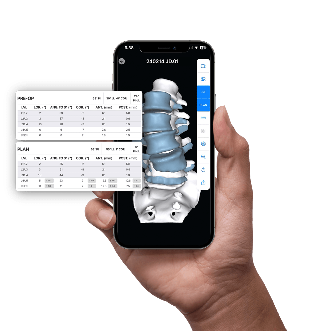
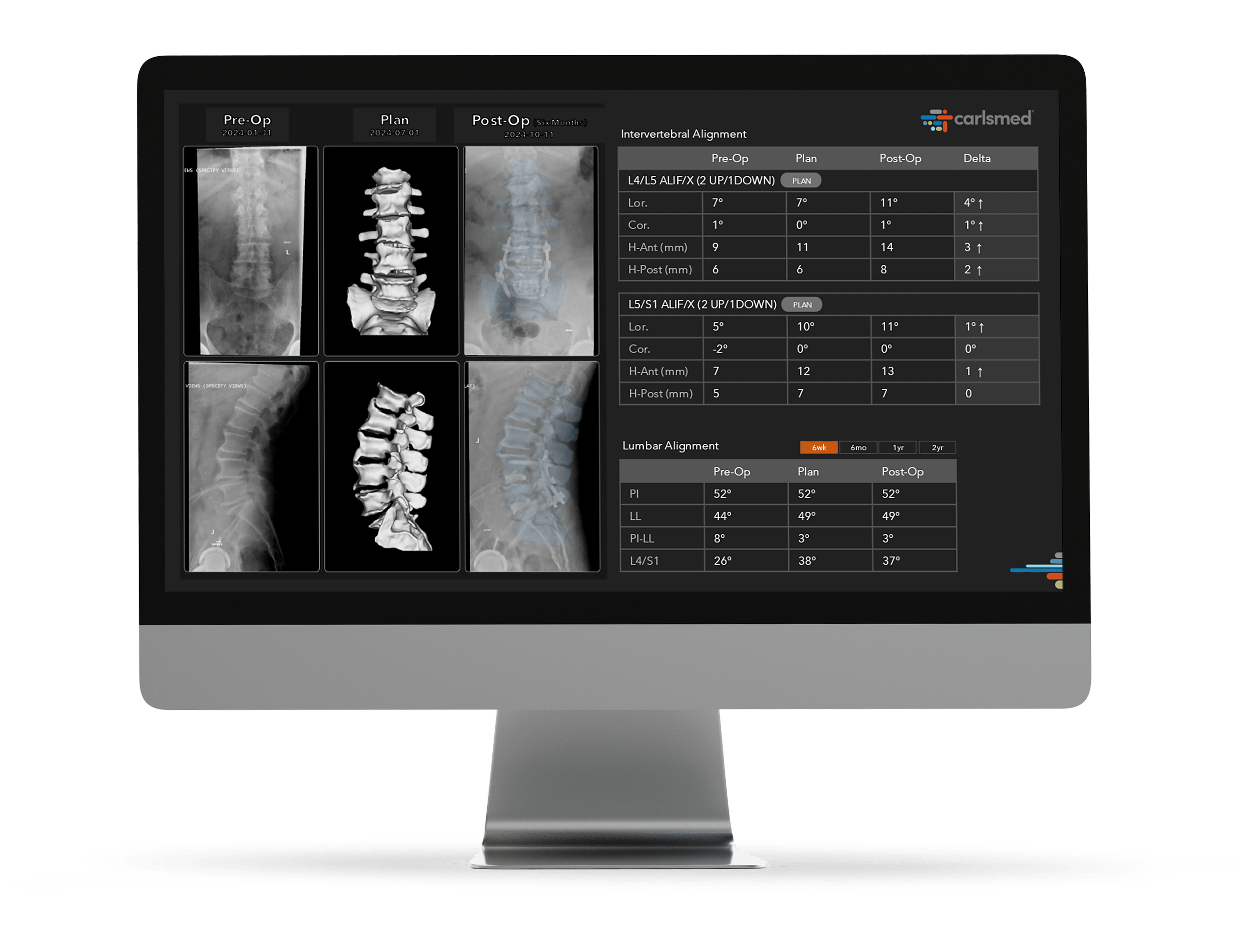
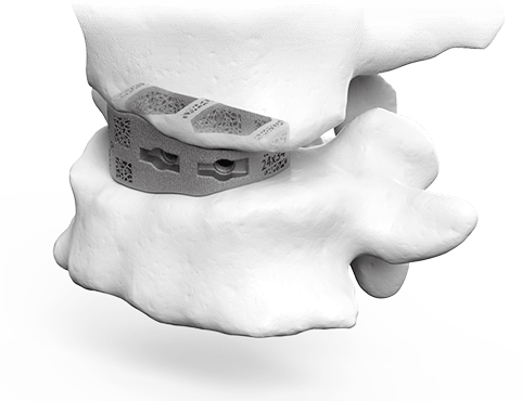
aprevo® was developed to help surgeons achieve the desired correction during surgery. Achieving the planned correction has been proven to reduce complications.5,6
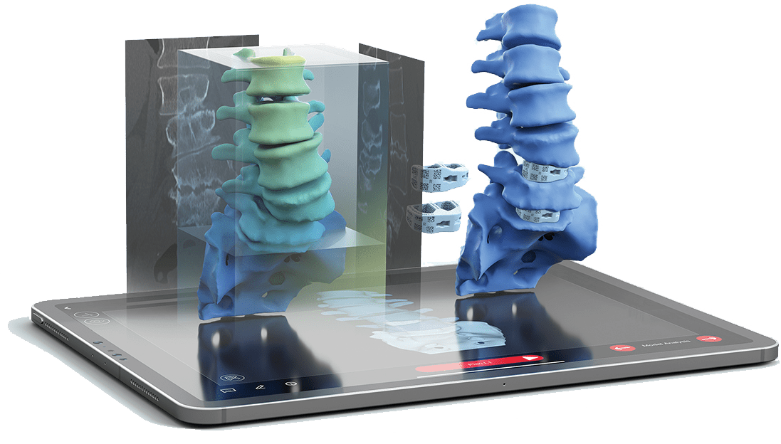
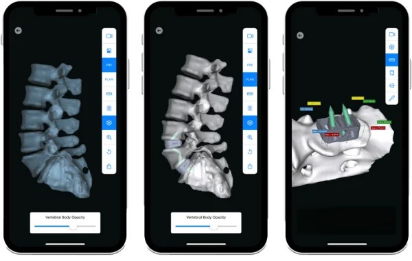
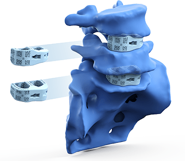
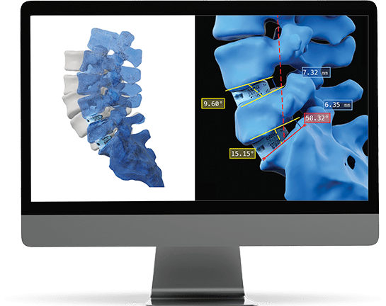
Carlsmed® provides a comprehensive Digital Solution for surgeons to inform, engage, and empower patients in the pre-operative decision-making process. Using proprietary algorithms, Carlsmed® combines the power of predictive analytics and prior outcomes data to develop personalized surgical plans and devices for each patient. Data is fed into the surgical planning loop to enable continuous learning, creating a closed loop around the surgeon.
For many patients with mild or moderate deformity, a short fusion construct may help in restoring alignment. Other patients may require a more significant correction. Carlsmed® develops the plan and devices based on your specific surgical goals for each patient. Just like you, we treat one patient at a time.
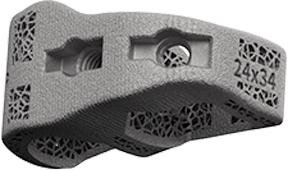
Personalized interbody devices are likely to provide for a more effective treatment vs the standard of care.7 By addressing the pathology and matching the anatomy of each individual patient, aprevo® may reduce complications, improve outcomes, and lower the cost of care. Improved outcomes lead to increased patient satisfaction.
Optimized surgical workflow and improved OR efficiency. aprevo® implants and associated instruments are provided sterile and packaged in one simple box. Traditional implant systems often require one or multiple instruments and implant trays that require sterilization. Our patient-specific model has been designed to minimize infection risk and reduce reprocessing costs. Patient-specific implants are also designed to streamline surgical workflow and may reduce operative time by reducing the need for trialing with traditional stock implant devices.
Call 1-866-aprevo1
1. Bess S, Line B, Fu MF, et al. The health impact of symptomatic adult spinal deformity: comparison of deformity types to United States population norms and chronic diseases. Spine. 2016; 41:E224–233.
2. Kalakoti P, Hendrickson N, Eisenberg J, Pugely A. Forecasting spinal deformity healthcare burden and operative utilization in the United States from 2015 to 2040: An epidemiological based ARIMA computation modeling. 2019 SRS Annual Meeting, Abstract 44
3. Moal B, Schwab F, Ames CP, et al. Radiographic outcomes of adult spinal deformity correction: A critical analysis of variability and failures across deformity patterns. Spine Deform. 2014 May;2(3):219-225.
4. Krol O, Passias PG, Lafage V, et al. Radiographic malalignment has a far greater impact on clinical outcomes than perioperative and postoperative complications in ASD surgery. 2021 SRS Annual Meeting, Abstract 18.
5. Rothenfluh DA, Mueller DA, Rotherfluh E, Min K. Pelvic incidence-lumbar lordosis mismatch predisposes to adjacent segment disease after lumbar spinal fusion. Eur Spine J. (2015) 24:1251–1258.
6. Tempel ZJ, Gandhoke GS, Bolinger BD, et al. The influence of pelvic incidence and lumbar lordosis mismatch on development of symptomatic adjacent level disease following single level transforaminal lumbar interbody fusion. Neurosurgery. 80:880–886, 2017.
7. FDA correspondence, 7-1-2020
8. Carlsmed data. NASS 2020
9. Ozgur BM, Gillard DM, Wood EE, et al. Can the use of a novel bone graft delivery system significantly increase the volume of bone graft material in a lumbar in situ cage, beyond volumes normally achieved via standard cage filling methodology? Results from a cadaveric pilot study. Interdisciplinary Neurosurgery. 14 (2018) 32–36.
10. Chatham LS, Patel VV, Yakacki CM, Carpenter RD. Interbody spacer material properties and design conformity for reducing subsidence during lumbar interbody fusion. J of Biomechanical Engineering. May 2017, Vol. 139 / 051005-1.
11. Fengbin, Yu, et al. Evaluation of a New Type of Titanium Mesh Cage versus the Traditional Titanium Mesh Cage for Single-Level, Anterior Cervical Corpectomy and Fusion. European Spine Journal, vol. 22. 12 (2013) 2891–2896.